Big biological datasets map life’s networks
Michael Snyder’s genes were telling him that he might be at increased risk for type 2 diabetes. The Stanford University geneticist wasn’t worried: He felt healthy and didn’t have a family history of the disease. But as he monitored other aspects of his own biological data over months and years, he saw that diabetes was indeed emerging, even though he showed no symptoms.

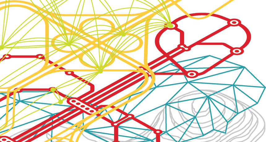
Snyder’s story illustrates the power of looking beyond the genome, the complete catalog of an organism’s genetic information. His tale turns the genome’s one-dimensional view into a multidimensional one. In many ways, a genome is like a paper map of the world. That map shows where the cities are. But it doesn’t say anything about which nations trade with each other, which towns have fierce football rivalries or which states will swing for a particular political candidate.
Open one of today’s digital maps, though, and numerous superimposed data sources give a whole lot of detailed, real-time information. With a few taps, Google Maps can show how to get across Boston at rush hour, offer alternate routes around traffic snarls and tell you where to pick up a pizza on the way.

Now, scientists like Snyder are developing these same sorts of tools for biology, with far-reaching consequences. To figure out what’s really happening within an organism — or within a particular organ or cell — researchers are linking the genome with large-scale data about the output of those genes at specific times, in specific places, in response to specific environmental pressures.
While the genome remains mostly stable over time, other “omes” change based on what genes are turned on and off at particular moments in particular places in the body. The proteome (all an organism’s proteins) and the metabolome (all the metabolites, or small molecules that are the outputs of biological processes) are two of several powerful datasets that become more informative when used together in a multi-omic approach. They show how that genomic instruction manual is actually being applied.
“The genome tells you what can happen,” says Oliver Fiehn, a biochemist at the University of California, Davis. The proteome and the metabolome can show what’s actually going on.

And just as city planners use data about traffic patterns to figure out where to widen roads and how to time stoplights, biologists can use those entwined networks to predict at a molecular level how individual organisms will respond under specific conditions.

By linking these layers and others to expand from genomics to multi-omics, scientists might be able to meet the goals of personalized medicine: to figure out, for example, what treatment a particular cancer patient will best respond to, based on the network dynamics responsible for a tumor. Or predict whether an experimental vaccine will work before moving into expensive clinical tests. Or help crops grow better during a drought.

And while many of those applications are still in the future, researchers are laying the groundwork right now.

“Biology is being done in a way that’s never been done before,” says Nitin Baliga, director of the Institute for Systems Biology in Seattle.

Data dump
Scientists have long studied how genes influence traits. Researchers have figured out important connections between genes and the proteins they encode and have scoured the genome for associations between particular genetic mutations and diseases. But a gene-by-gene view of the body is like trying to diagnose a citywide traffic problem by looking at just one backed-up intersection.

“There are so many places that a system can go awry,” Baliga says. When dozens of genes are working together, it’s tricky to tease out which one is misfiring in a particular instance.

Baliga is among a growing group of scientists who want to study life through a systems lens, because sometimes that traffic jam at one intersection is being caused by an out-of-sight accident three blocks away.

Such an approach is particularly useful for unraveling the complexities of diseases like cancer and diabetes. These conditions involve a tangled web of genes, paired with lifestyle factors and environmental conditions — Is she a smoker? How much does she exercise? — that influence when those various genes are turned on and off.
Reconstructing the tangled routes by which genes interact to influence the body is a slightly more complicated feat than mapping the best path from Tulsa to Tuscaloosa. For one thing, it requires serious computer power to gather, store and analyze all that data. The 3 billion chemical coding units that string together to form a person’s inventory of DNA, if entered into an Excel spreadsheet line-by-line, would stretch about 7,900 miles. The human proteome contains more than 30,000 distinct proteins that have been identified so far. And researchers have cataloged more than 40,000 different human metabolites, such lactic acid, ethanol and glucose.

Working with such big datasets can be expensive, too. Compiling the first human genome took 10 years and cost almost $3 billion. Now, the costs of collecting and analyzing all these datasets have come down, so it’s finally feasible to use them in tandem to answer big biological questions.

The important players
Scientists would like to understand the interplay between the genome and the proteome. Add in the metabolome. To make things more complex, there’s the epigenome — the chemical modifications to DNA that help control which genes are turned on and off — and the transcriptome, the full range of RNAs that translate DNA’s blueprints so they can be used to make proteins. It’s no surprise that mapping such a comprehensive network for any organism is still a distant goal.

For now, scientists tend to focus their multi-omic studies on a particular disease or question. Baliga wants to learn how tuberculosis — which sickens nearly 10 million people per year and kills 1.5 million — evades drugs within the body. Many strains of the TB bacterium are resistant to existing treatments or can tolerate them long enough to establish a strong foothold.

To learn how Mycobacterium tuberculosis mounts a defense against a drug, Baliga is first looking within the bacterium, identifying the genes, proteins and other molecules that interact as the microbe infects a host.

He collects different types of omic data from M. tuberculosis alone and when it’s in the presence of an antibiotic. His team recently focused on the microbe’s response to bedaquiline, a drug used to treat multidrug-resistant TB. Baliga measured the microbe’s transcriptome in the presence of different doses of bedaquiline and at different times after introducing the drug.

From this giant data dump, computer models helped narrow the focus to a smaller collection of genes, proteins and other molecules that changed under certain conditions. Visualization programs turned these mathematical outputs into maps that scientists could analyze.

About 1,100 genes behaved differently in the presence of bedaquiline, Baliga’s team reported in August in Nature Microbiology. Measurements of the RNA indicated that most of those genes became less active, but a few shifted into overdrive. The researchers suspected those hyperactive genes might be behind the resistance — playing off each other to create a smaller network within the larger tuberculosis response network.

But statistical analysis alone wasn’t enough to confirm the hunch. Correlation isn’t cause, Fiehn points out. Scientists need to figure out which of those changes actually matter.
That is, if you’re scanning millions of data points looking for variation, you’re going to find certain abnormalities that are due to chance and are unrelated to the disease or
question at hand. But starting from that smaller dataset of outputs that change, scientists can then test which players are actually important in the network and which belong on the sidelines. In animal models or petri dishes, scientists disable one gene at a time to see how it affects the proposed network.

“Systems biology has been able to generate these amazing hypotheses about how genes interact,” Baliga says. Testing them has historically been more challenging. But now, gene-editing technologies such as CRISPR/Cas9 (SN: 9/3/16, p. 22) allow scientists to more easily test these networks in living systems.

Baliga and his team edited the genome of M. tuberculosis, disabling the regulatory machinery responsible for some of the overactive genes. Sure enough, the drug worked better on the modified bacteria, the researchers reported.

Networking solutions
Once a network has been mapped, scientists can use it to predict (and maybe even prevent) illness.

Baliga’s team identified a second drug that works with bedaquiline. The drug turns off some of the regulators for the overactive tuberculosis gene network that was fighting off the bedaquiline. Using the second drug with bedaquiline made tuberculosis bacteria more vulnerable, pointing to a potential strategy for dealing with persistent infections.

Baliga’s group is also mapping networks in patients with glioblastoma, a particularly deadly type of brain tumor. In the August Cell Systems, the scientists described work designed to figure out why some patients respond to certain drugs while others don’t. The aim is to personalize treatments, to choose a drug that targets the particular network glitch that gives rise to that patient’s tumor, Baliga says. The drug might ramp up production of a protein that’s currently in short supply, or turn off a gene that’s mistakenly on. That same drug might be completely useless for another patient whose tumor developed through a different network error.

“Being able to do that systematically across all cancers, using networks — that has not happened yet,” Baliga says. But scientists have devised drug treatments to address individual mutations. And expanding that to a greater range of cancers in the future is not farfetched, he says.

Other scientists are using multi-omic approaches for preventive medicine, for example, to be more effective and efficient in vaccine development. Despite years of trying, scientists still haven’t created an HIV vaccine that can protect people against the virus, says Alan Aderem, a biologist at the Center for Infectious Disease Research in Seattle. Bringing a vaccine from test tube to market is costly and time-consuming. With a better understanding of how the networks of the body’s immune system respond to the disease, researchers could be more selective in the vaccine candidates that they invest time and money in developing.

“The real value of systems biology is that it’s predictive,” Aderem says. “If you could predict upfront if a vaccine would work, you’d not only save a huge amount of energy, you’d also save a huge amount of lives.”

Plant power
Multi-omics has perhaps received the most attention in the context of human health — but that’s also the realm where it’s hardest to piece together the omic layers. Because simpler organisms can be manipulated genetically, it’s easier to move from networks on a computer screen to real solutions.
For instance, some scientists are using multi-omic analysis to engineer networks that let crop plants thrive with less water. To turn carbon dioxide into sugar via photosynthesis, plants need a lot of water. But some desert plants make do with less. Most plants take in carbon dioxide through small pores in their leaves. Opening these pores can let water evaporate out. In the desert, where water is in short supply, some plants use a different network of chemical reactions to make energy: crassulacean acid metabolism, or CAM.

Plants that use CAM open pores in their leaves only during the cooler nighttime, when water is less likely to evaporate out. They store the CO2 they take in until daytime, when they close the pores and convert the CO2 into food.

“Our goal is to move this metabolic trick into crop plants,” says John Cushman, a biochemist at the University of Nevada, Reno. “In order to do that, you have to understand the complexity of all the enzymes and regulatory components associated with the circadian clock of the plant.”

Cushman’s team has collected vast omic datasets from CAM plants. He estimates that several hundred genes coordinate the process, turning each other on and off and producing proteins that interact with each other and influence other genes.

His team is trying to engineer the plant Arabidopsis (a weed often used for genetic experiments) to use CAM by inserting the relevant genes into the plant. If they can get it to work in this small lab plant, the researchers want to do the same in poplar trees to help grow biofuel sources in harsh environments not usually suitable for agriculture. Someday, Cushman hopes, the technology will help food crops grow better in arid climates.

Better than a hunch
So far, collecting and integrating omic data is largely restricted to the lab — the work is still expensive and time-consuming. But scientists like Snyder, who diagnosed himself with type 2 diabetes, think that someday people could regularly collect these sorts of data to track changes in their health over time.

The Stanford geneticist began by analyzing his own genome. Then he started measuring his proteome, metabolome and other omic data at regular intervals to develop what he termed his “personal omics profile.”

Certain mutations in his genome suggested that he was at risk for developing diabetes from the get-go. He was surprised, but didn’t take action. Nearly a year into his experiment, though, changes in his other omic data suggested that his body was no longer using glucose properly. His blood sugar was elevated and so was a type of hemoglobin that increases with uncontrolled diabetes, according to a report in Cell by Snyder and three dozen colleagues in 2012. Those aren’t new measures — they’re exactly what a doctor would test in someone with diabetes symptoms.
But Snyder had no symptoms. He had just had a nasty cold, which he now thinks may have triggered the onset of diabetes because he was already genetically at risk.

Because he spotted the changes early and his doctor confirmed he had diabetes, Snyder was able to fend off symptoms by changing his diet and exercise habits — which was reflected in the follow-up data he collected.

He monitored how other biological measurements (molecules that standard medical diagnostic tests might not even look at) changed with the onset of his diabetes and the lifestyle changes he made in response. That information might be valuable for doctors hoping to detect the onset of diabetes in patients before symptoms appear. So now his lab is tracking omic data from 100 people, most of whom have elevated blood sugar levels, but have not yet been diagnosed with diabetes. Snyder wants to see whether viral infections trigger diabetes in other people, or whether his case is an isolated incident.

Snyder is still tracking fluctuations in his own data over time, too. He thinks it’s a powerful tool for personalized medicine because it shows in real time how an individual responds to disease and other stressors.

“You’ll be able to start linking disease to treatment to outcomes,” Snyder says. “It becomes what I call data-driven medicine, instead of hunch-driven medicine.”
Making sense of this sort of data isn’t easy. “No story is the same,” Snyder says. “We see a lot of strange stuff that we don’t know how to interpret yet.”

But collecting detailed omic-level data about individuals over time could still be useful, he says. By collecting more data from healthy people, doctors and scientists could get a better sense of what’s normal. And tracking fluctuations in individual patients’ data over time can show what’s typical for that particular person. Snyder thinks that might be more valuable than knowing what’s normal for people in general. Someone monitoring these biological signs might notice molecular changes — as he did — before they cause troublesome physical symptoms.

For the multi-omic future that Snyder and others envision, scientists will need a greater ability to store and wrangle massive amounts of data than they possess today. And they’ll need ways to test the networks they uncover. But the potential payoff is enormous: A response map that can display the intertwining routes between genes and outcomes, as well as how distant red lights, speed bumps and construction zones play off each other to shift those paths.

Uranus moon count: 27 and rising

Two more teeny moons might be lurking around Uranus. That’s in addition to the 27 we already know about. Fluctuations in the density of two of the planet’s dark rings, seen in radio data from the 1986 flyby of the Voyager 2 spacecraft, could be caused by unseen moonlets, Robert Chancia and Matthew Hedman, astronomers at the University of Idaho in Moscow, report online October 9 at arXiv.org.

At probably just 4 to 14 kilometers wide, both moons would be very difficult to detect in Voyager 2 images, the researchers report. New observations with ground-based telescopes might have better luck.

New technique shows cells’ molecules in color

Electron microscopy is finally getting its Kodachrome moment.

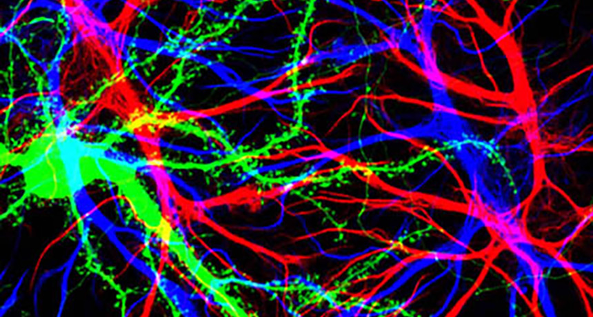
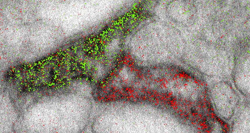
The high-powered scopes can now produce images that simultaneously highlight different molecules in different colors, scientists report online November 3 in Cell Chemical Biology. That’s helpful for researchers hoping to visualize the complex structures of cells or tissues — such as connections between brain cells, shown here.

Electron microscopes build black-and-white images by shooting beams of electrons at samples. Previously, scientists could add color by overlaying lower-resolution images from light microscopes. The new technique adds pizzazz without sacrificing image quality. It involves sequentially layering different metal ions on top of the sample. Each ion selectively latches onto a different target molecule. The electron beam interacts differently with each ion, yielding signature wave shapes that can be converted into colors. The researchers used the coloring technique to show that two brain cells called astrocytes (the edge of one shown in green, the other in red) could link up to the same message-sending junction between nerve cells.

Website turns Alzheimer’s research into a game

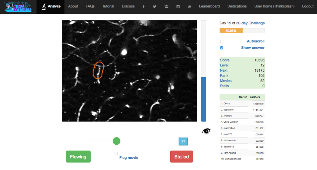
Traffic jams in the brain’s blood supply may play a role in Alzheimer’s disease. A new online game turns people at home into amateur traffic cops. This policing, which involves spotting hard-to-see sluggishness in tiny capillaries in mice, may ultimately help scientists better understand, and perhaps even treat, Alzheimer’s, a devastating disorder that affects over 5 million Americans.

The science behind the game, called Stall Catchers, comes from Cornell University. Chris Schaffer, Nozomi Nishimura and colleagues found that mice designed to exhibit symptoms similar to Alzheimer’s have more blocked blood vessels in their brains than regular mice. That difference can deprive the brain of sustenance and may be a key to understanding how Alzheimer’s damages the brain, the researchers suspect.
But finding congested capillaries is a slog. Computers haven’t been up to snuff, and experts could spend an entire year analyzing the thousands of microscope images needed to amass enough data to explore links between Alzheimer’s and blocked vessels. “I thought, if we could change that, it would be tremendous,” says Pietro Michelucci, director of the Human Computation Institute in Fairfax, Va. The institute is a nonprofit organization that runs the EyesOnALZ program, which aims to crowdsource Alzheimer’s research.

That’s where StallCatchers.com comes in. The website asks players to sift through short black-and-white videos of real mouse brains, on the prowl for blocked blood vessels. In the videos, moving blood appears white. But stationary black segments that appear between two white segments signal trouble — a stall. Players rack up points and ascend levels as they classify vessels. With practice, the task gets easier. And people who suffer from performance anxiety shouldn’t fret; each video will be scrutinized by multiple users to get the final verdict. With a little help from the crowd, “not only do [researchers] get answers faster, but they can ask more questions,” Michelucci says.

So far, nearly 1,000 users have played Stall Catchers, Michelucci says. Those players are beginning to generate data that will let researchers see how good these amateur traffic cops are. With luck, their eyes will help unstall the fight against Alzheimer’s.

Heartburn drugs may raise stroke risk

NEW ORLEANS — Popular heartburn drugs — already under investigation for possible links to dementia, kidney and heart problems (SN: 6/11/16, p. 8) — have a new health concern to add to the list. An analysis of almost 250,000 medical records in Denmark has found an association with stroke.

Researchers from the Danish Heart Foundation in Copenhagen studied patients undergoing gastric endoscopy from 1997 to 2012. About 9,500 of all patients studied suffered from ischemic strokes, which occur when a blood clot blocks a blood vessel in the brain.

Overall, the risk of stroke was 21 percent higher in patients taking a proton pump inhibitor, a drug that relieves heartburn, the researchers reported November 15 during the American Heart Association’s annual meeting. While those patients also tended to be older and sicker to start with, the level of risk was associated with dose, the researchers found. People taking the lowest drug doses (between 10 and 20 milligrams a day, depending on the drug) did not have a higher risk. At the highest doses, though, Prevacid (more than 60 mg/day) carried a 30 percent higher risk and Protonix (more than 80 mg/day) a 94 percent higher risk. For Prilosec and Nexium, stroke risk fell within that range.

Introduced in the 1980s, proton pump inhibitors are available in both prescription and over-the-counter forms. While they are valuable drugs, “their use has been increasing rapidly,” says lead author Thomas Sehested, adding that people often take them for too long, or without a clear reason. Before taking them, he says, “patients need a conversation with their doctor to see if they really need these drugs.”

Plant-eating mammals sport bigger bellies than meat eaters

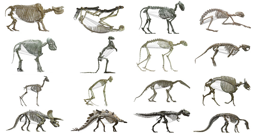
These skeletons are spilling their guts about the size of the body cavity that housed these animals’ stomach and intestines.

Using digital 3-D scans of mounted skeletons, researchers estimated the body cavity volume in 126 species. Of the 76 mammal species, plant eaters had bigger bellies; their relative torso volumes were about 1.5 times as large as those of carnivores, researchers report online November 4 in the Journal of Anatomy.

The study is the first to quantitatively test the long-held idea that herbivores have bigger torsos, says Marcus Clauss of the University of Zurich. Plant eaters are thought to need extra space for complex systems that digest a leafy diet.
Surprisingly, Clauss and colleagues didn’t find the same pattern in nonavian dinosaurs, birds or reptiles, but the researchers had fewer skeletons to compare. Of the 27 dinosaurs, for example, only four were carnivores.

Still, the research suggests that in tetrapods — four-limbed vertebrates — only mammalian herbivores have larger body cavities, raising questions about why that might be evolutionarily. “Everybody goes crazy about the long neck or the strange things” on an animal’s head, Clauss says. But few scientists have focused on the torso’s frame and how diet helps sculpt it over time. “This study emphasizes that the torso is an important part of overall body shape.”

Health official calls on neuroscience to fight mental illness

SAN DIEGO — Society’s record for protecting public health has been pretty good in the developed world, not so much in developing countries. That disparity has long been recognized.

But there’s another disparity in society’s approach to public health — the divide between attention to traditional diseases and the resources devoted to mental disorders.

“When it comes to mental health, all countries are developing countries,” says Shekhar Saxena, director of the World Health Organization’s department of Mental Health and Substance Abuse. Despite a breadth of scope and depth of impact exceeding that of many more highly publicized diseases, mental illness has long been regarded as a second-class medical concern. And modern medicine’s success at diagnosing, treating and curing many other diseases has not been duplicated for major mental disorders.

Saxena thinks that neuroscience research can help. He sees an opportunity for progress through increased interdisciplinary collaboration between neuroscience and mental health researchers.

“The collaboration seems to be improving, but much more is needed and not only in a few countries, but all countries,” he said November 12 at the annual meeting of the Society for Neuroscience.

By almost any measure, mental health disorders impose an enormous societal burden. Worldwide, direct and indirect costs of mental disorders exceed $2.5 trillion yearly, Saxena said — projected to reach $6 trillion by 2030. Mental illnesses also disable and kill in large numbers: Global suicides per year total over 800,000. “Indeed, it is a hidden epidemic,” Saxena said. That’s more deaths than from breast cancer and probably more from malaria, according to a new comprehensive analysis of global mortality.

Mental illnesses encompass a wide range of disorders, from autism and Alzheimer’s disease to substance abuse and schizophrenia. Saxena acknowledged that there have been advances in the scientific understanding of these diseases, but not nearly enough. No easily used diagnostic test is available for most of them. And no new class of drugs for treating major mental disorders has appeared in the last 20 years, with the possible exception of dementia.

“We need increased investment in research, increased public, private and philanthropic investments,” Saxena said. “We need increased connection of research with public health gains.”
He called on the community of neuroscientists to establish a “grand challenge” to researchers to address these concerns.

“There have been many grand challenges in neurosciences — perhaps we need one for finding out mental health interventions,” he said.

Many neuroscientists are, of course, aware of the important link between their research and mental health. Some progress is being made. One promising avenue of work focuses on synapses, the junctures through which nerve cells in the brain communicate. Typically synapses form where axons, the long neuronal extensions that transmit signals, connect with dendrites, the neurons’ message-receiving branches. Most of the axon-dendrite connections occur at small growths called dendritic spines that protrude from the dendrite surface.

Several sessions at the neuroscience meeting described new work showing ways in which dendritic spines may be involved in mental disorders. One session focused on the protein actin, a prime structural component of the spines.

Actin activity depends on a complicated chain of chemical reactions inside a cell. In mice, blocking a key link in that chain reduces the number of spines in the front part of the brain, Scott Soderling of Duke University School of Medicine reported. Those mice then exhibited symptoms reminiscent of schizophrenia in people.

It seems that losing spines in the frontal cortex alters nerve cell connections there; with spine shortages, some axons link directly to the dendrite shaft. Bypassing spines, which play a filtering role, can intensify signals sent to the ventral tegmental area, which in turn may send signals that increase production of the chemical messenger dopamine in another brain region, the striatum. “We think that this is actually what is driving the elevated dopamine levels” in disorders such as schizophrenia, Soderling said.

Some antipsychotic drugs (known as neuroleptics) for treating schizophrenia work by blocking sites of dopamine action in the striatum. But such drugs do nothing about the loss of spines that initiated the problem.

“These neuroleptics largely treat these symptoms, but they’re not a cure,” Soderling said. “We think that this is good evidence … for the idea that these drugs are treating a downstream consequence of a primary insult that’s occurring elsewhere in the brain.”

This insight from neuroscience — that antipsychotics treat downstream symptoms, not the problem at its source — may help the search for better treatments.

Soderling suggests that many other mental disorders may have their roots in problems with actin in dendritic spines. Another speaker at the neuroscience meeting, Haruo Kasai of the University of Tokyo, emphasized how fluctuations in the numbers of spines, related to actin activity, could play a role in autism.

Such results from neuroscience should be of great value to fighting mental disorders. But science alone won’t enable the discovery of effective treatments without a broader scope of scientific investigation of mental illness as a global problem. Too much of research to date focuses on too small a portion of the worldwide population. As Saxena noted, more than 90 percent of scientific studies on mental illness are from — and about — high-income countries.

“We are ignoring a very large number of people living in this world,” he said. “And this can be, and is, a real impediment to science. If we don’t know what is happening in the brains of the majority of the people living in this world, can we really advance science in the best possible manner? Can we still say that we know the human brain? And at least my answer would be: No.”

Year in review: ‘Three-parent baby’ technique raises hope and concern

A “three-parent baby” was born in April, the world’s first reported birth from a controversial technique designed to prevent mitochondrial diseases from passing from mother to child.

“As far as we can tell, the baby is normal and free of disease,” says Andrew R. La Barbera, chief scientific officer of the American Society for Reproductive Medicine. “This demonstrates that, in point of fact, the procedure works.”

The baby boy carries DNA not only from his mother and father but also from an egg donor, raising both safety and ethical concerns. In particular, people worry that alterations of the genetic makeup of future generations won’t stop with preventing diseases but could lead to genetically enhanced “designer babies.”
Opponents, such as Marcy Darnovsky, executive director of the Center for Genetics and Society in Berkeley, Calif., are also worried that the technique hasn’t been fully tested. “We wish the baby and family well, and hope the baby stays healthy,” Darnovsky says. “But I have a lot of concerns about this child and about future efforts to use these techniques before they’ve been shown to be safe.”

About one in 4,000 children are born with dysfunctional mitochondria. These energy-generating organelles are inherited from the mother and have their own DNA. Mutations in some of the 37 mitochondrial genes can lead to fatal diseases, often affecting energy-hungry organs such as the brain and muscles. Because there is no cure or effective treatment for many mitochondrial diseases, the recent birth has been heralded as a sign of new hope for affected families.

Even if women don’t have mitochondrial diseases themselves, they can pass the diseases to their children if their egg cells contain large numbers of defective mitochondria. The mother of the recent three-parent baby had previously had two children who died of Leigh syndrome, a mitochondrial disease that affects the nervous system and eventually prevents a person from breathing.

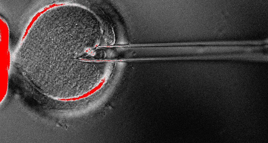
Fertility doctor John Zhang of the New Hope Fertility Center in New York City and colleagues performed what’s called a spindle transfer to put all the chromosomes from the mother’s egg into a donor egg that contained healthy mitochondria but had been emptied of its chromosomes (SN Online: 10/18/16). The egg was then fertilized with sperm and implanted in the mother.

Cell swap
A baby boy born in April has DNA from three people. To produce the embryo, researchers transferred the chromosomes from the mother’s egg into a donor egg with healthy mitochondria. The technique is called “spindle transfer” for the cellular structure that segregates the chromosomes.

“It’s very important that they follow up,” to monitor the child’s long-term health, says Shoukhrat Mitalipov, a mitochondrial biologist at Oregon Health & Science University in Portland. Mitalipov pioneered the spindle transfer technique in monkeys (SN: 9/26/09, p. 8). Even a small number of defective mitochondria carried over from the mother’s egg may replicate and cause problems later on, he and other scientists have found (SN: 6/25/16, p. 8; SN Online: 11/30/16).

Zhang reported that just 1.6 percent of the baby boy’s mitochondrial DNA came from his mother (SN Online: 10/19/16). Mitalipov notes, however, that doctors can’t know from sampling a few types of tissue whether other tissues have different levels of mitochondrial carryover. What’s more, levels of mutant mitochondria may change as the child grows.

Mitalipov supports research on the technique but says it should be done in carefully controlled clinical trials. Results of a mouse study published in July suggest that mismatches between the parents’ nuclear DNA and the donor mitochondrial DNA could affect metabolism and aging (SN: 8/6/16, p. 8). Those effects could show up years or decades after birth.

The baby boy born in April is technically not the first three-parent baby. At least two children born in the late 1990s carry mitochondrial DNA from a donor. Those two and 15 other children were born to mothers who had a small amount of cytoplasm — the gelatinous fluid that fills cells and holds mitochondria — from a donor egg injected into their own eggs in an effort to improve results of in vitro fertilization. No major health problems have been reported, but the studies were abandoned because of ethical concerns, lack of funding and the difficulties in obtaining newly required permits.

La Barbera disputes the term “three-parent baby” entirely. “A person’s essence as a human being comes from their nuclear genetic material, not their mitochondrial genetic material,” La Barbera says. Children who are born after mitochondrial transfer procedures have only two parents, he contends.

Zhang drew fire for going to Mexico to perform the procedure. Congress currently bars the U.S. Food and Drug Administration from reviewing applications to make heritable changes in human embryos, which includes the spindle transfer technique. A panel of experts said in February that it is ethical to make three-parent baby boys (SN Online: 2/3/16), a provision that would prevent future generations from inheriting the donor mitochondria. Because mothers pass mitochondria on to their babies but fathers usually do not, technically baby boys born through this technique don’t carry an inheritable modification in their DNA.

Clinics in the United Kingdom can legally perform the procedures, but none have been reported yet. A panel of experts there recommended November 30 that clinical studies could move ahead, so more babies may be born in 2017.

New blood tests can detect prions

A new blood test can detect even tiny amounts of infectious proteins called prions, two new studies show.

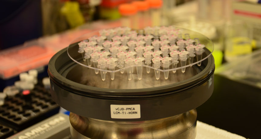
Incurable prion diseases, such as mad cow disease (BSE) in cattle and variant Creutzfeldt-Jakob disease (vCJD) in people, result from a normal brain protein called PrP twisting into a disease-causing “prion” shape that kills nerve cells in the brain. As many as 30,000 people in the United Kingdom may be carriers of prions that cause vCJD, presumably picked up by eating BSE-tainted beef. Health officials worry infected people could unwittingly pass prions to others through blood transfusions. Four such cases have already been recorded. But until now, there has been no way to screen blood for the infectious proteins.
In the test, described December 21 in Science Translational Medicine, magnetic nanobeads coated with plasminogen — a protein that prions grab onto — trap prions. Washing the beads gets rid of the rest of the substances in the blood. Researchers then add normal PrP to the beads. If any prions are stuck to the beads, the infectious proteins will convert PrP to the prion form, which will also stick to the beads. After many rounds, the researchers could amplify the signal enough to detect vCJD prions in all the people in the studies known to have the disease.

No healthy people or people with other degenerative brain diseases (including Alzheimer’s and Parkinson’s) in either study had evidence of the infectious proteins in their blood. And only one of 83 people with a sporadic form of Creutzfeld-Jakob disease tested positive. Those results indicate that the test is specific to the vCJD prion form, so a different test is needed to detect the sporadic disease.

In two cases, researchers detected prions in frozen blood samples collected 31 months and 16 months before people developed vCJD symptoms.

Dark matter still missing

Chalk up one more loss for physicists searching for dark matter. Scientists with the XENON100 experiment have largely ruled out another experiment’s controversial claim of detecting dark matter.

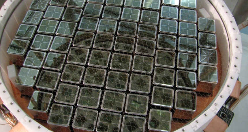
XENON100, located in Italy’s Gran Sasso National Laboratory, aims to directly detect particles of dark matter — the unknown substance that scientists believe makes up the bulk of matter in the cosmos (SN: 11/12/16, p. 14).

In their new analysis, published online January 3 at arXiv.org, XENON100 scientists looked for an annual variation in the rate of blips in their detector, a tank filled with 161 kilograms of liquid xenon. Such a signal could be a hallmark of Earth’s motion through a prevailing wind of dark matter particles as the planet makes its yearly jaunt around the sun. Another dark matter experiment at Gran Sasso, DAMA/LIBRA, claims to have found strong evidence of a yearly modulation, but other experiments have failed to replicate the result.
Scientists combed over four years of data for events that could be caused by dark matter interacting with electrons in XENON100. The researchers found no evidence of an annual cycle, contradicting DAMA’s claim.

Dark matter optimists can still cling to a caveat, though: DAMA uses a different detection material, composed of sodium iodide crystals rather than xenon. That might explain the difference between the two experiments. Future experiments will attempt to replicate DAMA’s result using the same material.